 To my home page
 To School of Physics Home Page
More info from dnj@physics.unimelb.edu.au
By Jacinta den Besten and David N. Jamieson
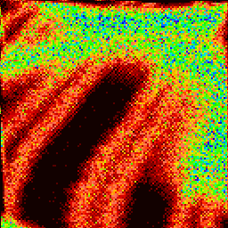
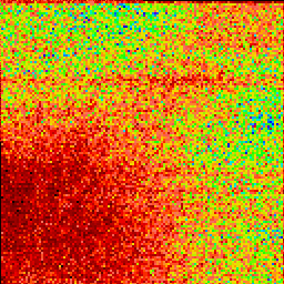
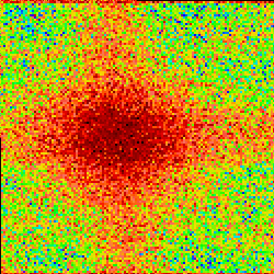
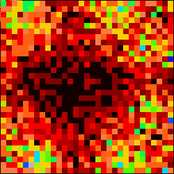
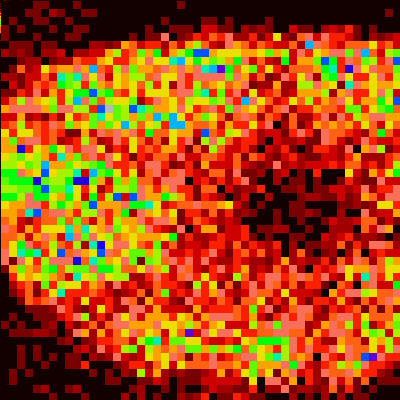
These beam rocking images were obtained with MP2 using two pre-lens scan coils and a 3 MeV H beam.
Experimental patterns:
Grid scan in full beam rocking mode. What does it mean? The beam spot appeared to be tracing a square on the sample.
Ni in the 100 direction
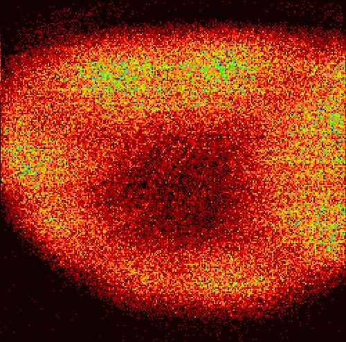
Ni in the 100 direction, maximum possble scan (note vignetting caused by beam hitting beam tube). Scan size was +/- 0.9 degrees.
So we can do it for 100 Si as well. Scan size was +/-0.45 degrees.
And for tiny amounts of epi-SiGe on the surface of the previous Si crystal. Same maximum scan size as before.
And indeed hexagonal crystals like sapphire work just fine too.
More info from dnj@physics.unimelb.edu.au